Project Is Supported by Over $2.4 Million Grant from NIH

Electrical and Computer Engineering Prof. Hengyong Yu, right, with Ph.D. student Yongshun Xu.
05/12/2023
By Edwin L. Aguirre
Heart disease is the leading cause of death for men and women in America, according to the U.S. Centers for Disease Control and Prevention.
One person dies every 34 seconds in the United States from cardiovascular disease, the CDC reports, and this costs the country about $229 billion each year in health care services, medicines and lost productivity due to disability or death.
A team of researchers from UMass Lowell, Rensselaer Polytechnic Institute and Vanderbilt University Medical Center led by UML Electrical and Computer Engineering Prof. Hengyong Yu is developing technology that would greatly improve cardiac CT scans, which doctors currently use to diagnose cardiovascular diseases, so that timely, life-saving treatment and preventive measures can be implemented.
Aside from Yu, other key investigators include Electrical and Computer Engineering Prof. Yan Luo (UML), Prof. Ge Wang (RPI) and Prof. J. Jeffrey Carr (VUMC).
The project is supported by a four-year grant worth more than $2.4 million from the National Institutes of Health’s National Institute of Biomedical Imaging and Bioengineering.
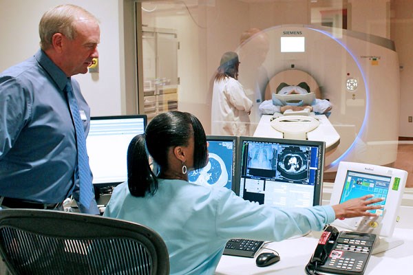 Image by National Institutes of Health
Cardiac CT scans are an important tool that doctors use to diagnose cardiovascular diseases in patients.
Yu and his co-investigators are developing a new image-reconstruction algorithm based on artificial intelligence (AI) that would effectively “freeze” the beating heart in CT images within a brief, 60-millisecond time window (one twentieth of a heartbeat).
“This would eliminate the blurring movement of the coronary arteries in X-ray images and help doctors analyze plaque buildup on the walls of the arteries, which is the main cause of heart attacks,” Yu says.
“Moreover, our method will not require patients to hold their breath during the CT exam and will eliminate the need to use beta-blocker drugs to slow down the patients’ heart rates,” he says.
According to Yu, the team’s AI-based computational framework would radically improve the image quality of existing CT scanners and would benefit patients who suffer from tachycardia (rapid heartbeat) and arrhythmia (irregular heartbeat) that commonly occur in older adults, many of whom experience atrial fibrillation (rapid, irregular heart rhythm).
“Our project will combine two innovative image-processing algorithms – compressed sensing and deep learning – to reconstruct cardiac CT images at very high resolution and with lower radiation exposure to patients compared to traditional CT scans,” Yu notes.
He says their technique could allow them to help build powerful, low-cost cardiac CT scanners, and possibly retrofit older models to perform cardiac CT exams.
“Our algorithm could dramatically expand the capability of these systems, allowing higher-quality cardiac CT scans in many underprivileged communities worldwide.”
Assisting Yu in the lab research is Yongshun Xu, a fourth-year electrical engineering doctorate student.
“I’m actively recruiting more postdocs and graduate students,” says Yu. “I hope to get two postdocs and two Ph.D. students to join the project this fall.”